Rat Model for Seminal Vesicle Occlusion (SVO)
- Product No.DSI837Ra01
- Organism SpeciesRattus norvegicus (Rat) Same name, Different species.
- Prototype SpeciesHuman
- SourceBilateral seminal vesicle ligation method
- Model Animal StrainsSPF SD rats (male,8 weeks)
- Modeling GroupingRandomly divided into groups: Control group, Model group, Positive drug group and Test drug group
- Modeling Period2 weeks
- Modeling MethodConstruction of seminal vesicle occlusion rat model
The group of seminal vesicle occlusion
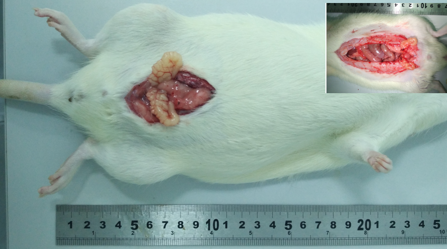
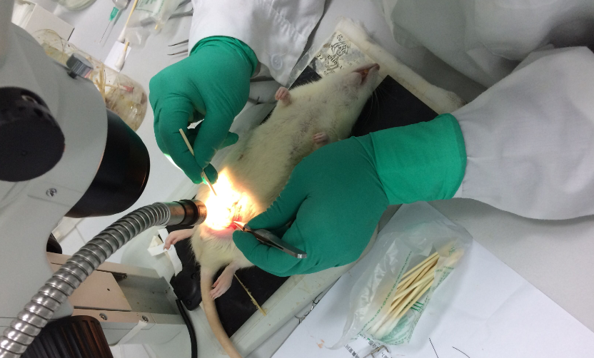
50 male Wistar rats aged of 8 weeks were selected, with environment temperature at 20-25℃, humidity 40-70%. After a week of adaptive ordinary feeding, 25 rats selected at random served as the sham group, the others as the seminal vesicle occlusion group were anaesthetized by pentobarbital sodium, cut their belly for 15-20mm to expose seminal vesicle, preformed the operation of bilateral seminal vesicle ligation by a silk ring, then the seminal vesicle was returned back in suit, and the wound was seamed. For the sham group, their belly was cut for 15-20mm to expose seminal vesicle, then it was returned back in suit, and the wound was seamed. - ApplicationsDisease Model
- Downloadn/a
- UOM Each case
- FOB
US$ 200
For more details, please contact local distributors!
Model Evaluation
1. The effect of seminal vesicle occlusion on the rat’s visceral coefficient
1.1 Observation of seminal vesicle size
The rats were fasted for 12 hours before killed, and weighed in each group. The rats were killed after narcotized with barbital sodium. Then the rats were laparotomized to expose their seminal vesicles, and the size of seminal vesicle was observed in each group .
1.2 Detection of Viscera weight
Took out bilateral testis, epididymis, seminal vesicle, peeled off the surrounding adipose tissue, washed with cold saline, removed blood, imbibed water with filter paper, weighted with electronic balance.
Calculate viscera index: viscera index = the weight of viscera/ the weight of rat body
2.The Secretary function of seminal vesical
2.1 secretion volume
Took out the seminal vesicle. After weighing, the seminal vesicle fluid was squeezed into the test tube and weighted by a electronic balance.
2.2 Fructose concentration
Added chymotrypsin aqueous solution into the seminal vesicle fluid, detected fructose concentration by a fructopyranose test kits.
The result showed that in occlusion rat model, the seminal vesicle was smaller, secretion of seminal vesicle reduced, fructose concentration of seminal vesicle fluid reduced.
3. the effect of seminal vesicle occlusion on the rat’s sexual function :
The experiment was began for 1 hours after the light was dark. 10 female rats were injected 30ug estradiol benzoate at 24 hours before the experiment. And 10 female rats were injected 50ug luteosterone at 4 hours before the experiment. Mating test was preformed in darker environments. At first, put male rats into the cage (50cm×30cm×20cm) for 10 min to suit themselves, then put a female estrous rat into the gate, immediately observed and recorded the sexual behavior of male rats. The parameters were below:
1.(mount latency, ML): the time from when caged with female rats to the first frequency
2.(mount frequency, MF): the amount of frequency within 2 hours whether there was an intromission.
3.(intromission latency, IL): the time from when caged with female rats to the first intromission
4.(intromission frequency, IF): the amount of intromission within 2 hours
5.(ejaculation latency, EL): the time from the first intromission to ejaculation
6.(ejaculation frequency, EF): the ejaculation frequency of rats within 2 hours
7.(rat number of ejaculation behavior, EBN): the number of rats with a ejaculation behavior
8.(hit rate, HR): the rate of intromission to mount latency
The next day, observed vagina of female rats whether there was a vaginal suppository. If found, recorded a successful mating . If not, a test paper was inserted into vagina and examined under the microscope, sperm was found to be a successful mating.
Pathological Results
After the seminal vesicle fluid was completely squeezed out, added formaldehyde into one side of the seminal vesicle to prepare parafin section. Then stained by HE, observed and recorded under microscope.
There were many fold in the seminal vesicle, the surface was pseudostratified columnar epithelium, the cells contained secretory granules, fat droplets and lipofuscin, and the sexual function was impaired.
Cytokines Level
Statistical Analysis
SPSS software is used for statistical analysis, measurement data to mean ± standard deviation (x ±s), using t test and single factor analysis of variance for group comparison, P<0.05 indicates there was a significant difference, P<0.01 indicates there are very significant differences.
GIVEAWAYS
INCREMENT SERVICES
-
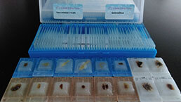 Tissue/Sections Customized Service
-
 Serums Customized Service
-
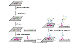 Immunohistochemistry (IHC) Experiment Service
-
 Small Animal In Vivo Imaging Experiment Service
-
 Small Animal Micro CT Imaging Experiment Service
-
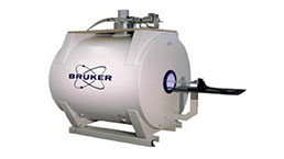 Small Animal MRI Imaging Experiment Service
-
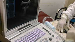 Small Animal Ultrasound Imaging Experiment Service
-
 Transmission Electron Microscopy (TEM) Experiment Service
-
 Scanning Electron Microscope (SEM) Experiment Service
-
 Learning and Memory Behavioral Experiment Service
-
 Anxiety and Depression Behavioral Experiment Service
-
 Drug Addiction Behavioral Experiment Service
-
 Pain Behavioral Experiment Service
-
 Neuropsychiatric Disorder Behavioral Experiment Service
-
 Fatigue Behavioral Experiment Service
-
 Nitric Oxide Assay Kit (A012)
-
 Nitric Oxide Assay Kit (A013-2)
-
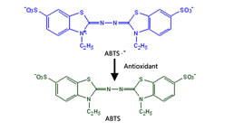 Total Anti-Oxidative Capability Assay Kit(A015-2)
-
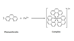 Total Anti-Oxidative Capability Assay Kit (A015-1)
-
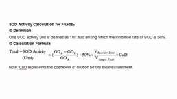 Superoxide Dismutase Assay Kit
-
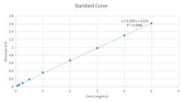 Fructose Assay Kit (A085)
-
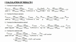 Citric Acid Assay Kit (A128 )
-
 Catalase Assay Kit
-
 Malondialdehyde Assay Kit
-
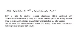 Glutathione S-Transferase Assay Kit
-
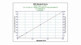 Microscale Reduced Glutathione assay kit
-
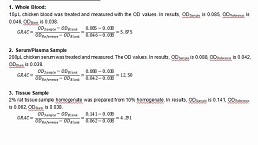 Glutathione Reductase Activity Coefficient Assay Kit
-
 Angiotensin Converting Enzyme Kit
-
 Glutathione Peroxidase (GSH-PX) Assay Kit
-
 Cloud-Clone Multiplex assay kits
| Catalog No. | Related products for research use of Rattus norvegicus (Rat) Organism species | Applications (RESEARCH USE ONLY!) |
| DSI837Ra01 | Rat Model for Seminal Vesicle Occlusion (SVO) | Disease Model |


