
Packages (Simulation)
Reagent Preparation
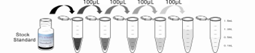
Image (I)
-
 Results demonstration
Image (II)
-
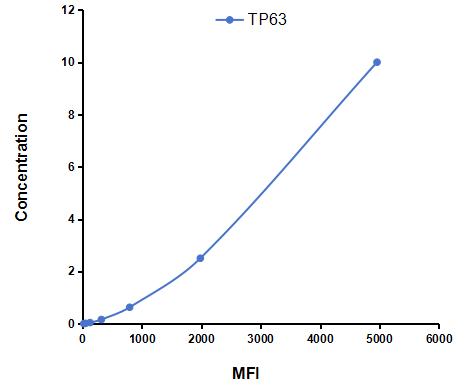 Typical Standard Curve
Quality Guarantee

Instruction Manual
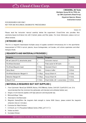
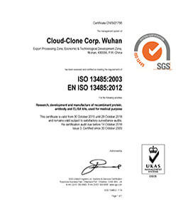
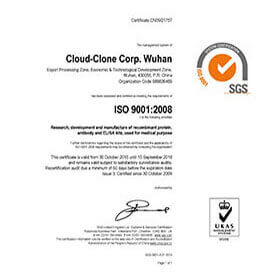
Certificate
Multiplex Assay Kit for Tumor Protein P63 (TP63) ,etc. by CBA (Cytometric Bead Array)
P63; TP73L; CUSP; EEC3; KET; LMS; OFC8; RHS; SHFM4; TP53CP; Tumor Protein p53-Competing Protein; Chronic ulcerative stomatitis protein; Keratinocyte transcription factor
- Product No.CMC805Mu
- Organism SpeciesMus musculus (Mouse) Same name, Different species.
- Sample TypeTissue homogenates and other biological fluids.
- Test MethodDouble-antibody Sandwich
- Assay Length4h
- Detection Range0.0098-10ng/mL
- SensitivityThe minimum detectable dose of this kit is typically less than 0.0033ng/mL.
- DownloadInstruction Manual
- UOM 48T96T
- FOB
US$ 605
US$ 864
For more details, please contact local distributors!
Specificity
This assay has high sensitivity and excellent specificity for detection of Tumor Protein P63 (TP63) ,etc. by CBA (Cytometric Bead Array).
No significant cross-reactivity or interference between Tumor Protein P63 (TP63) ,etc. by CBA (Cytometric Bead Array) and analogues was observed.
Recovery
Matrices listed below were spiked with certain level of recombinant Tumor Protein P63 (TP63) ,etc. by CBA (Cytometric Bead Array) and the recovery rates were calculated by comparing the measured value to the expected amount of Tumor Protein P63 (TP63) ,etc. by CBA (Cytometric Bead Array) in samples.
| Matrix | Recovery range (%) | Average(%) |
| serum(n=5) | 84-103 | 99 |
| EDTA plasma(n=5) | 89-104 | 97 |
| heparin plasma(n=5) | 92-101 | 95 |
Precision
Intra-assay Precision (Precision within an assay): 3 samples with low, middle and high level Tumor Protein P63 (TP63) ,etc. by CBA (Cytometric Bead Array) were tested 20 times on one plate, respectively.
Inter-assay Precision (Precision between assays): 3 samples with low, middle and high level Tumor Protein P63 (TP63) ,etc. by CBA (Cytometric Bead Array) were tested on 3 different plates, 8 replicates in each plate.
CV(%) = SD/meanX100
Intra-Assay: CV<10%
Inter-Assay: CV<12%
Linearity
The linearity of the kit was assayed by testing samples spiked with appropriate concentration of Tumor Protein P63 (TP63) ,etc. by CBA (Cytometric Bead Array) and their serial dilutions. The results were demonstrated by the percentage of calculated concentration to the expected.
| Sample | 1:2 | 1:4 | 1:8 | 1:16 |
| serum(n=5) | 95-103% | 87-96% | 81-92% | 89-104% |
| EDTA plasma(n=5) | 92-105% | 85-104% | 83-92% | 79-101% |
| heparin plasma(n=5) | 94-101% | 88-96% | 79-101% | 79-102% |
Stability
The stability of kit is determined by the loss rate of activity. The loss rate of this kit is less than 5% within the expiration date under appropriate storage condition.
To minimize extra influence on the performance, operation procedures and lab conditions, especially room temperature, air humidity, incubator temperature should be strictly controlled. It is also strongly suggested that the whole assay is performed by the same operator from the beginning to the end.
Reagents and materials provided
| Reagents | Quantity | Reagents | Quantity |
Assay procedure summary
1. Add 200μL analysis buffer solution to each well of the plate for pre-wetting;
2. Preparation of standards, reagents and samples before the experiment;
3. Add 100μL standard or sample to each well, add 10μL magnetic beads, and incubate 120 minutes protect from light at 25°C on shaker;
4. Remove liquid on magnetic frame, add 100μL prepared Detection Reagent A. Incubate 60 minutes protect from light at 25°C on shaker;
5. Remove liquid on magnetic frame, add 100μL prepared Detection Reagent B, and incubate 30 minutes protect from light at 25°C on shaker;
6. Wash plate once on magnetic frame;
7. Add 100μL sheath fluid, swirl for 10 minutes, read on the machine.
GIVEAWAYS
INCREMENT SERVICES
| Catalog No. | Related products for research use of Mus musculus (Mouse) Organism species | Applications (RESEARCH USE ONLY!) |
| RPC805Mu01 | Recombinant Tumor Protein P63 (TP63) | Positive Control; Immunogen; SDS-PAGE; WB. |
| RPC805Mu02 | Recombinant Tumor Protein P63 (TP63) | Positive Control; Immunogen; SDS-PAGE; WB. |
| PAC805Mu01 | Polyclonal Antibody to Tumor Protein P63 (TP63) | WB; IHC; ICC; IP. |
| PAC805Mu02 | Polyclonal Antibody to Tumor Protein P63 (TP63) | WB; IHC; ICC; IP. |
| SEC805Mu | ELISA Kit for Tumor Protein P63 (TP63) | Enzyme-linked immunosorbent assay for Antigen Detection. |
| CMC805Mu | Multiplex Assay Kit for Tumor Protein P63 (TP63) ,etc. by CBA (Cytometric Bead Array) | FLIA Kit for Antigen Detection. |
| LMC805Mu | Multiplex Assay Kit for Tumor Protein P63 (TP63) ,etc. by FLIA (Flow Luminescence Immunoassay) | FLIA Kit for Antigen Detection. |