ELISA to detect autophagy, next hot spot?
Autophagy, proposed by Ashford and Porter in 1962 after they found the "eat themselves" phenomenon within cells, refers to part of cytoplasm from no ribosome attached district of rough endoplasmic reticulum, which was embed by bilayer membrane. The cytoplasm and the organelles, and protein and the other components need degradation formed autophagosome. The autophagosome and lysosome merged and formed autophagosome-lysosome complex, the complex will degrade all the embed content. In this way, the metabolism demand of the cells will be met and some organelles will be updated . Currently autophagy is one of the most hottest topics in life science area following apoptosis ,according to literature record in pubmed, about 1500 papers are reported before 2006. The total amount of related paper published from 2007 to September, 2010 reached about 4,400.
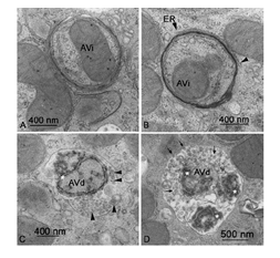
However, how to detect the autophagy, is still a big problem for the researchers. The gold standard for detecting autophagy is to directly observe the phagophore under the electron microscope. As shown in Figure 1, we can see various forms of phagophore. This method are not used widely because of the limit of equipment and the skill to operate it. Therefore, LC3 antibodies can also be used in Western Blot, according to the ratio of LC3-II/I, the phase of autophagy can be evaluated. When autophagy happens, a short peptide LC3-I will be removed from cytoplasic LC3 by enzymatic hydrolysis. Then LC3-I and PE combine into (autophagosomes) membrane type (that is, LC3-II).

Fig1. The forms of phagosome in different phrase under the electron microscope (From: Madame Curie Bioscience Database)
In addition, fluorescence proteins such as GFP-LC3 and mRFP-GFP-LC3 are also widely used in the detection of autophagy. GFP-LC3 can be expressed by transfection or infection in target cells, then autophagy can be evaluted by fluorescence microscopy according to the quantity and strength of GFP.
As enzyme-linked immunosorbent assay technology developed in recent years, researchers found that the ELISA method can also be easily used to evaluate autophagy. The principle of the method is to detect autophagy-related gene. autophagy associated gene, ATG, as its name suggests, refers to genes directly participate or regulated the process of Autophagy. So far,there are 16 ATG which were discovered, named from ATG1 to ATG16. Among them, ATG8 is also known as LC3, and ATG6 is known as Beclin-1.
Based on it, Cloud-Clone Corp. has developed a series of ATG proteins assay kits, including the ATG12detection kit (SEL224Hu), ATG16L1 detection kit (SEN942Hu), ATG5 detection kit(SEL221Hu), ATG7 detection kit (SEL222Hu), BECN1 detection kit (SEJ557Hu), ATG16L1 detection kit (SEN942Hu), ATG16L1 detection kit (SEN942Hu) and so on. These products provide a good platform for researchers in the field of autophagy.
Compared with other methods to detect autophagy, ELISA test has many advantages. For example, it is more convenient and time saving. This is no need for the expensive equipment such as electron microscopy or fluorescence microscopy. Moreover it can detect multiple samples simultaneously. Therefore, ELISA method provides great support for the meahanism study of autophagy .
For more related products, please visit www.cloud-clone.us.
