
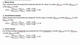
Packages (Simulation)
Image (II)
-
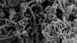
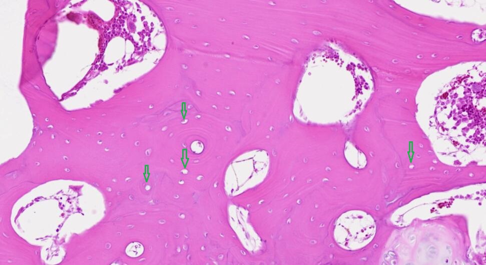 Fig.HE staining of femur on Model rabbit ,Arrow: Bone lacunae
-
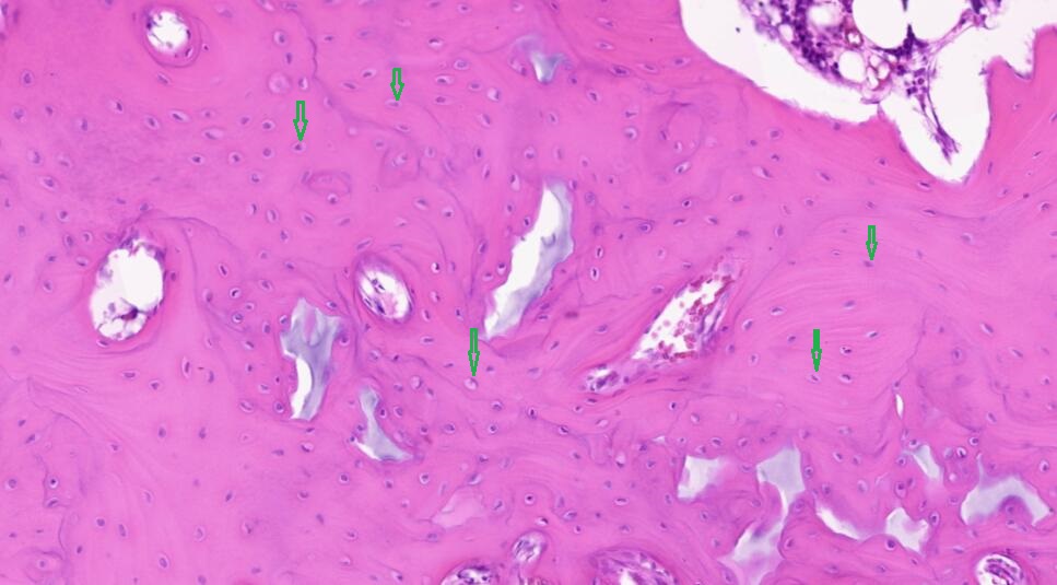 Fig.HE staining of femur on Control rabbit, Arrow: Bone lacunae
Quality Guarantee

Certificate


Rabbit Model for Avascular Necrosis (AN)
Avascular Necrosis of femoral head;
- Product No.DSI589Rb02
- Organism SpeciesOryctolagus cuniculus (Rabbit) Same name, Different species.
- Prototype SpeciesHuman
- SourceSteroid-induced Avascular Necrosis of femur, SANFH
- Model Animal StrainsBig ears white rabbit, healthy, male, weight 2.0-2.5kg.
- Modeling GroupingRandomly divided into two group: Control group and Model group.
- Modeling Period4-6 weeks
- Modeling Method1. Animal grouping: Japanese male big ear rabbits were randomly divided into 2 groups: Control group and Model group.
2. Animal modeling: After a week of adaptive feeding, start the modeling by Hexijing modeling method.
Model group: Inject prednisolone acetate (8mg/kg) on the right gluteus muscle of the rabbit, twice a week for 6 weeks.
Control group: Inject equal normal saline on the right gluteus muscle of the rabbit, twice a week for 6 weeks.
Prophylactic injection of penicillin (80,000 units/kg) on the left gluteus muscle were provided to all animals, twice a week for 6 weeks. - ApplicationsDiesase model
- Downloadn/a
- UOM Each case
- FOB
US$ 500
For more details, please contact local distributors!
Model Evaluation
Model identification: After 6 weeks, take caput femoris and collect aorta abdominalis blood. Then kill rabbits in blank group and model group using air embolization method.
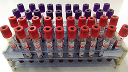
Blood specimen: Forbidden diet for 24 hours before sampling, collect aorta abdominalis blood under general anesthesia into a glass tube, refrigerate, centrifuge, remove upper serum into EP tube, save at -20℃.
Caput femoris specimen: After blood collection of abdominal aorta, take the right caput femoris in an aseptic condition. Dissect along the coronal plane and place in 4% paraformaldehyde, electron microscope fixation solution and liquid nitrogen in turn.
Observe appearance and quality of caput femoris, color and shape of arthrodial cartilage. Feel the hardness of the bone using a knife.
Pathological Results
Using HE staining, observe changes in form, structure and quantity of trabeculae, osteocyte, pulp cavity and hematopoietic cells under a light microscope to determine whether the model is successful or not.
Randomly select 10 fields under high power lens, count 50 bone lacuna in each field, calculate blank bone lacuna rate.
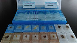
HE staining contains sampling, decalcification, embedding, slicing and staining.
Cytokines Level
1. Determine ALP in serum of each group by chemical colorimetry, determine TG by oxidase method according to the instructions.
2. Detect protein expression of p-ERK1, p-ERK2, p-JNK and p-P38 by Western blotting.
Statistical Analysis
SPSS software is used for statistical analysis, measurement data to mean ± standard deviation (x ±s), using t test and single factor analysis of variance for group comparison, P<0.05 indicates there was a significant difference, P<0.01 indicates there are very significant differences.
GIVEAWAYS
INCREMENT SERVICES
-
 Tissue/Sections Customized Service
-
 Serums Customized Service
-
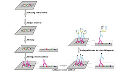 Immunohistochemistry (IHC) Experiment Service
-
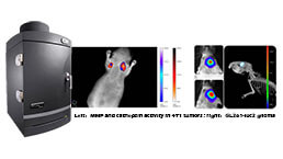 Small Animal In Vivo Imaging Experiment Service
-
 Small Animal Micro CT Imaging Experiment Service
-
 Small Animal MRI Imaging Experiment Service
-
 Small Animal Ultrasound Imaging Experiment Service
-
 Transmission Electron Microscopy (TEM) Experiment Service
-
 Scanning Electron Microscope (SEM) Experiment Service
-
 Learning and Memory Behavioral Experiment Service
-
 Anxiety and Depression Behavioral Experiment Service
-
 Drug Addiction Behavioral Experiment Service
-
 Pain Behavioral Experiment Service
-
 Neuropsychiatric Disorder Behavioral Experiment Service
-
 Fatigue Behavioral Experiment Service
-
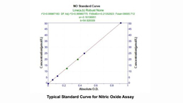
 Nitric Oxide Assay Kit (A012)
-
 Nitric Oxide Assay Kit (A013-2)
-
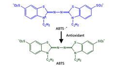 Total Anti-Oxidative Capability Assay Kit(A015-2)
-
 Total Anti-Oxidative Capability Assay Kit (A015-1)
-
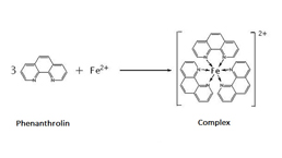
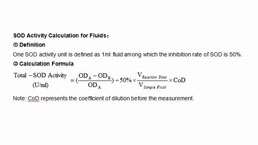 Superoxide Dismutase Assay Kit
-
 Fructose Assay Kit (A085)
-
 Citric Acid Assay Kit (A128 )
-
 Catalase Assay Kit
-
 Malondialdehyde Assay Kit
-
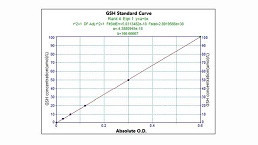
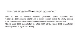 Glutathione S-Transferase Assay Kit
-
 Microscale Reduced Glutathione assay kit
-
 Glutathione Reductase Activity Coefficient Assay Kit
-
 Angiotensin Converting Enzyme Kit
-
 Glutathione Peroxidase (GSH-PX) Assay Kit
-
 Cloud-Clone Multiplex assay kits
| Catalog No. | Related products for research use of Oryctolagus cuniculus (Rabbit) Organism species | Applications (RESEARCH USE ONLY!) |
| DSI589Rb01 | Rabbit Model for Avascular Necrosis (AN) | n/a |
| DSI589Rb02 | Rabbit Model for Avascular Necrosis (AN) | Diesase model |