
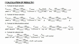
Packages (Simulation)
Image (II)
-
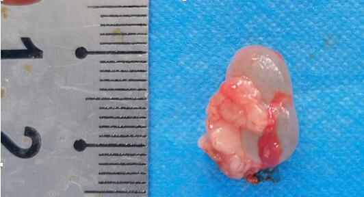 Fig. The lesion of the transplanted endometrium
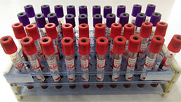
Quality Guarantee

Certificate


Rat Model for Endometriosis (EM)
Endometriosis uterina
- Product No.DSI543Ra01
- Organism SpeciesRattus norvegicus (Rat) Same name, Different species.
- Prototype SpeciesHuman
- SourceTransplantation of allograft uterine endometrium
- Model Animal StrainsSprague Dawley(SD)rat , healthy, female, 6-8W, body weight 200g-220g
- Modeling GroupingRandomly divided into six group: Control group, Model group, Positive drug group and Test drug group (three doses)
- Modeling Period4w
- Modeling Method1. Randomly select one rat as a donor and euthanize it by cervical spondylolysis. Fix it in a supine position on the operating table, disinfect the abdomen with alcohol, and then cut open the abdominal cavity along the midline. Find the Y-shaped uterus, carefully pull out the left and right sides of the uterus, and carefully separate the bilateral uterine mesentery.
2. Detach the uterine horn and ovaries, cut the left and right uterus separately at the Y-shaped uterine bifurcation, and place them in a sterile curved disc containing physiological saline. Repeatedly rinse the blood stains on the surface of the uterus to further separate the remaining adherent uterine mesentery. Place two uterine horns in two sterile curved discs and cut them directly into equal sizes of approximately 1mm × 1mm fragments were suspended in physiological saline.
3. The uterus of the donor mice are implanted in the abdominal cavity of the recipient rat in a ratio of 1/4. One donor rat is supplied with endometrium to four recipient rat, and the tissue fragments in the curved disc were suspended with physiological saline.
4. Randomly select a rat as the recipient, and take the left lower abdomen at the midline below the umbilicus as the injection point. Inject physiological saline containing uterine tissue fragments into the recipient's abdominal cavity with a numbered needle, along with uterine tissue fragments, and then place it in the rat cage. - ApplicationsUsed to study the occurrence process of endometriosis And drug intervention and treatment plans
- Downloadn/a
- UOM Each case
- FOB
US$ 250
For more details, please contact local distributors!
Model Evaluation
1. 4 weeks after surgery, ectopic lesion tissues are taken and observed under an optical microscope for morphology and histopathological results.
2. The model group rats have four regular estrous cycles, namely pre estrus, estrus, post estrus, and inter estrus, with each cycle lasting 4-5 days.
3. In the model group of rats, bulbous protrusions can be seen in the abdomen with naked eyes, showing cystic solidity and no activity. After cutting the skin, transparent bulbous cystic accretion can be seen, with a soft texture and clear liquid accumulation inside the capsule. Ectopic lesion tissue can be seen growing into a cavity like structure at low magnification under an optical microscope, while endometrial epithelial cells, stromal cells, and a small amount of glandular formation can be seen at high magnification. The histopathological manifestations are similar to those of human EMS.
Pathological Results
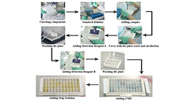
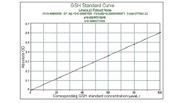
Cytokines Level
Statistical Analysis
SPSS software is used for statistical analysis, measurement data to mean ± standard deviation (x ±s), using t test and single factor analysis of variance for group comparison, P<0.05 indicates there was a significant difference, P<0.01 indicates there are very significant differences.
GIVEAWAYS
INCREMENT SERVICES
-
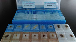 Tissue/Sections Customized Service
-
 Serums Customized Service
-
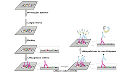 Immunohistochemistry (IHC) Experiment Service
-
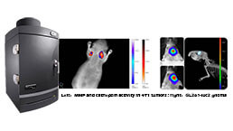 Small Animal In Vivo Imaging Experiment Service
-
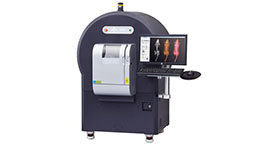 Small Animal Micro CT Imaging Experiment Service
-
 Small Animal MRI Imaging Experiment Service
-
 Small Animal Ultrasound Imaging Experiment Service
-
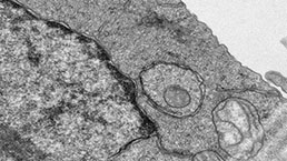 Transmission Electron Microscopy (TEM) Experiment Service
-
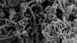 Scanning Electron Microscope (SEM) Experiment Service
-
 Learning and Memory Behavioral Experiment Service
-
 Anxiety and Depression Behavioral Experiment Service
-
 Drug Addiction Behavioral Experiment Service
-
 Pain Behavioral Experiment Service
-
 Neuropsychiatric Disorder Behavioral Experiment Service
-
 Fatigue Behavioral Experiment Service
-
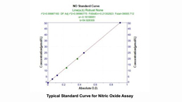
 Nitric Oxide Assay Kit (A012)
-
 Nitric Oxide Assay Kit (A013-2)
-
 Total Anti-Oxidative Capability Assay Kit(A015-2)
-
 Total Anti-Oxidative Capability Assay Kit (A015-1)
-
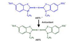
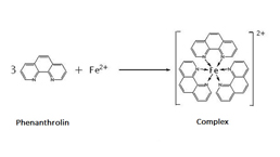
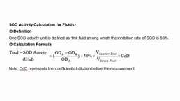 Superoxide Dismutase Assay Kit
-
 Fructose Assay Kit (A085)
-
 Citric Acid Assay Kit (A128 )
-
 Catalase Assay Kit
-
 Malondialdehyde Assay Kit
-
 Glutathione S-Transferase Assay Kit
-
 Microscale Reduced Glutathione assay kit
-
 Glutathione Reductase Activity Coefficient Assay Kit
-
 Angiotensin Converting Enzyme Kit
-
 Glutathione Peroxidase (GSH-PX) Assay Kit
-
 Cloud-Clone Multiplex assay kits
| Catalog No. | Related products for research use of Rattus norvegicus (Rat) Organism species | Applications (RESEARCH USE ONLY!) |
| DSI543Ra01 | Rat Model for Endometriosis (EM) | Used to study the occurrence process of endometriosis And drug intervention and treatment plans |