
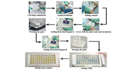
Packages (Simulation)
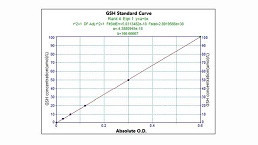
Image (I)
-
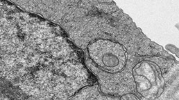
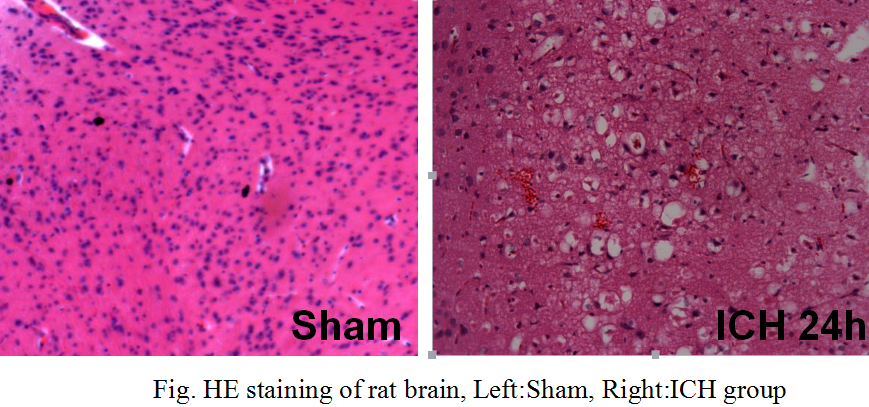 Fig. HE staining of rat brain, Left:Sham, Right:ICH group
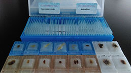
Image (II)
-
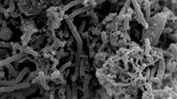
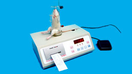
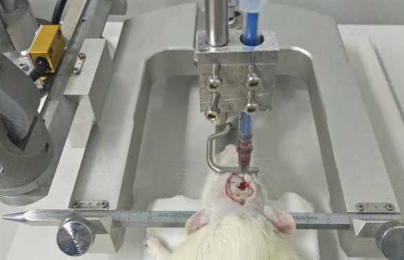 Fig. Autologous blood Microinjection into caudate nucleus
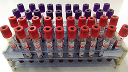
-
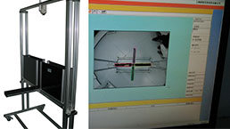
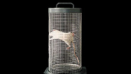
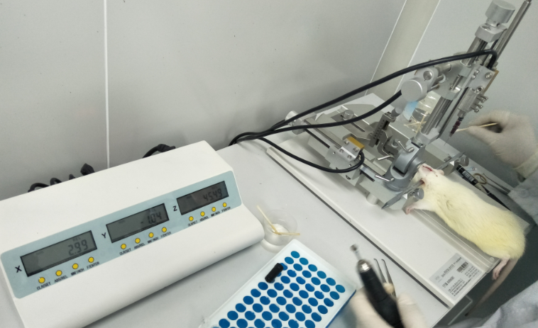 Fig. Stereotaxic localization of rat brain
Quality Guarantee

Certificate


Rat Model for Intracerebral Hematoma (IH)
- Product No.DSI686Ra01
- Organism SpeciesRattus norvegicus (Rat) Same name, Different species.
- Prototype SpeciesHuman
- SourceAutologous blood Microinjection into caudate nucleus
- Model Animal StrainsWistar Rats(SPF), healthy, male, body weight 250~300g.
- Modeling GroupingRandomly divided into six group: Control group, Model group, Positive drug group and three Test drug group.
- Modeling Period1~2 weeks
- Modeling Method1. Rats were anesthetized with intraperitoneal injection of 10% chloral hydrate (350mg/kg). Using a syringe to collect 100ul blood in the tail.Rest for 5 minutes until the blood is complete coagulation.
2.The rats were fixed on the stereotaxic apparatus.Make a median longitudinal incision, 30% hydrogen peroxide corrosion of periosteum exposed anterior fontanel and puncture point. Target localization in the anterior fontanelle before 1mm, the left side of the center line at 3mm, the outer surface of skull 6mm. At the insertion point, the skull is drilled with a diameter of 1mm round hole to the surface of the dura mater.
3.Inject the clot into the target with a micro syringe(injection rate 20ul/min), Suture the skin of the head, the rats were placed in a heating pad to maintain the temperature of 37 to 0.5 degrees Celsius to revive, normal feeding.
4.In the sham operation group, blood was taken from the tail, but no autologous blood was injected. - ApplicationsDisease Model
- Downloadn/a
- UOM Each case
- FOB
US$ 300
For more details, please contact local distributors!
Model Evaluation
1.HE staining observation:
HE staining in Sham group, no significant pathological changes were observed. The brain cells were arranged closely and neatly. 24 hours after the surgery, perihematomal tissue edema and necrosis, hyperchromatic nuclei shrinkage, edema, degeneration and necrosis of neurons, and accompanied by a large number of red blood cells, infiltration of inflammatory cells, nerve cells arranged in disorder, loose, reticular structure, cell gap increases.
2.Fluorescence quantitative PCR (Q-PCR) detection of brain tissue samples:
Expression content change of Caspase-3 and c-IAP-1 in the brain tissue around the hematoma (inhibitor of apoptosis protein) were measured.
Pathological Results
Cytokines Level
Statistical Analysis
SPSS software is used for statistical analysis, measurement data to mean ± standard deviation (x ±s), using t test and single factor analysis of variance for group comparison, P<0.05 indicates there was a significant difference, P<0.01 indicates there are very significant differences.
GIVEAWAYS
INCREMENT SERVICES
-
 Tissue/Sections Customized Service
-
 Serums Customized Service
-
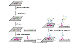 Immunohistochemistry (IHC) Experiment Service
-
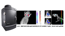 Small Animal In Vivo Imaging Experiment Service
-
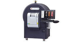 Small Animal Micro CT Imaging Experiment Service
-
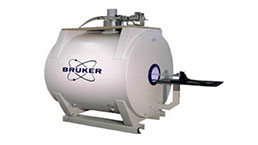 Small Animal MRI Imaging Experiment Service
-
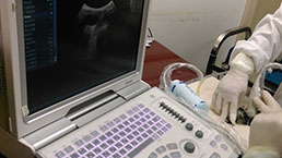 Small Animal Ultrasound Imaging Experiment Service
-
 Transmission Electron Microscopy (TEM) Experiment Service
-
 Scanning Electron Microscope (SEM) Experiment Service
-
 Learning and Memory Behavioral Experiment Service
-
 Anxiety and Depression Behavioral Experiment Service
-
 Drug Addiction Behavioral Experiment Service
-
 Pain Behavioral Experiment Service
-
 Neuropsychiatric Disorder Behavioral Experiment Service
-
 Fatigue Behavioral Experiment Service
-
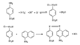 Nitric Oxide Assay Kit (A012)
-
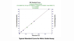 Nitric Oxide Assay Kit (A013-2)
-
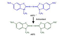 Total Anti-Oxidative Capability Assay Kit(A015-2)
-
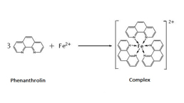 Total Anti-Oxidative Capability Assay Kit (A015-1)
-
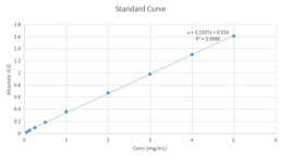
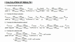
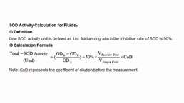 Superoxide Dismutase Assay Kit
-
 Fructose Assay Kit (A085)
-
 Citric Acid Assay Kit (A128 )
-
 Catalase Assay Kit
-
 Malondialdehyde Assay Kit
-
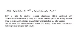 Glutathione S-Transferase Assay Kit
-
 Microscale Reduced Glutathione assay kit
-
 Glutathione Reductase Activity Coefficient Assay Kit
-
 Angiotensin Converting Enzyme Kit
-
 Glutathione Peroxidase (GSH-PX) Assay Kit
-
 Cloud-Clone Multiplex assay kits
| Catalog No. | Related products for research use of Rattus norvegicus (Rat) Organism species | Applications (RESEARCH USE ONLY!) |
| DSI686Ra01 | Rat Model for Intracerebral Hematoma (IH) | Disease Model |