
Packages (Simulation)
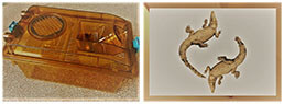
Image (II)
-
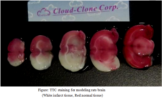 Fig. TTC staining for modeling rats brain
Quality Guarantee

Certificate


Rat Model for Cerebral Ischemia (CI)
Brain Ischemia; Cerebrovascular Ischemia; MCAO
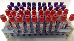
- Product No.DSI523Ra01
- Organism SpeciesRattus norvegicus (Rat) Same name, Different species.
- Prototype SpeciesHuman
- SourceIschemia-Reperfusion with MCAO
- Model Animal StrainsSD rats (SPF level), healthy, male, body weight 250g-300g.
- Modeling GroupingRandomly divided into six group: Control group, Model group, Positive drug group and Test drug group (three doses)
- Modeling Period24 hours, 3 days or 7 days
- Modeling Method1. 15% chloral hydrate to anesthetize rats, neck skin preparation, disinfection, insert the anus temperature probe, maintain the temperature within 37±0.5℃.
2. Incision of the middle of the neck, exposure the right common carotid artery, internal carotid artery and external carotid artery. Using 6-0 silk (at 4mm distance from the common carotid artery bifurcation ) to ligate the external carotid artery at the far end of the heart, pierces another 6-0 silk in the external carotid artery , and hit a slipknot near the bifurcation of the common carotid artery.
3. Use arterial clamps to clamp the common carotid artery. Make a small cut on the external carotid artery (3mm distance from bifurcation of common carotid artery ), a root tip treated 0.33 mm diameter nylon line from the cut insertion into the internal carotid artery, and inward into the middle cerebral artery, nylon line insertion depth distance carotid artery bifurcation about 16±1mm.
4. Ischemia 90 mins and pull the suture, a 6-0 silk ligates artery proximal, 3-0 silk suture wounds on the neck, povidone iodine disinfection wound, rat put in a heating pad, and feed on constant temperature raising box after rats awake.
5. 24hrs after operation, neurological function score was evaluated, and then the rats were injected intraperitoneally with 15% hydrate of chlorine aldehyde, and the brain was stained with TTC and pathological staining. - ApplicationsUsed to study the pathogenesis of cerebral infarction, pathophysiological process and thrombolytic therapy.
- Downloadn/a
- UOM Each case
- FOB
US$ 300
For more details, please contact local distributors!
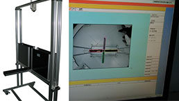
Model Evaluation
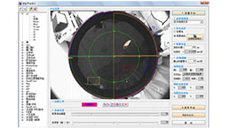
1.Neurological dysfunction score
Longa and Bederson's 5 point systems,make a score on rats waked from anaesthesia 24 hours later.
0 points: no nerve injury symptoms
1 points can not fully extend the forepaw
2 points: to the opposite side
3 points: to the side dump
4 points: can not walk spontaneously, loss of consciousness
2.TTC staining
Take the brain and store at -20℃,1% TTC (W/V), 37℃ water bath for TTC dissolved, the frozen brain tissue slices, placed in 10ml TTC solution, 37℃,10min. Normal brain tissue staining was bright red, and the infarct area was pale.
Pathological Results
Take the brain, 4% poly Formaldehyde Solution fixed, after dehydration of sucrose solution, the OCT embedded to make the frozen section (slice 10um), Nissl staining and the staining resluts are used for the evaluation of infarct size.
Cytokines Level
Statistical Analysis
SPSS software is used for statistical analysis, measurement data to mean ± standard deviation (x ±s), using t test and single factor analysis of variance for group comparison, P<0.05 indicates there was a significant difference, P<0.01 indicates there are very significant differences.
GIVEAWAYS
INCREMENT SERVICES
-
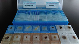 Tissue/Sections Customized Service
-
 Serums Customized Service
-
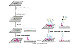 Immunohistochemistry (IHC) Experiment Service
-
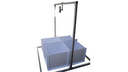
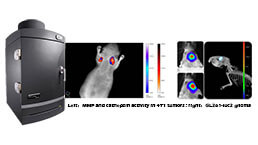 Small Animal In Vivo Imaging Experiment Service
-
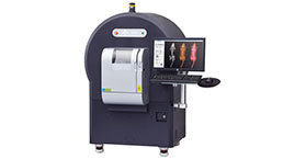 Small Animal Micro CT Imaging Experiment Service
-
 Small Animal MRI Imaging Experiment Service
-
 Small Animal Ultrasound Imaging Experiment Service
-
 Transmission Electron Microscopy (TEM) Experiment Service
-
 Scanning Electron Microscope (SEM) Experiment Service
-
 Learning and Memory Behavioral Experiment Service
-
 Anxiety and Depression Behavioral Experiment Service
-
 Drug Addiction Behavioral Experiment Service
-
 Pain Behavioral Experiment Service
-
 Neuropsychiatric Disorder Behavioral Experiment Service
-
 Fatigue Behavioral Experiment Service
-
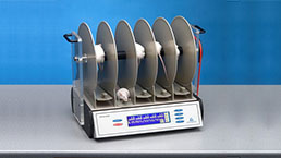
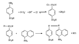 Nitric Oxide Assay Kit (A012)
-
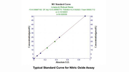 Nitric Oxide Assay Kit (A013-2)
-
 Total Anti-Oxidative Capability Assay Kit(A015-2)
-
 Total Anti-Oxidative Capability Assay Kit (A015-1)
-
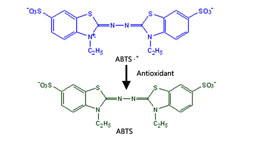
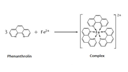
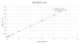
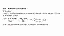 Superoxide Dismutase Assay Kit
-
 Fructose Assay Kit (A085)
-
 Citric Acid Assay Kit (A128 )
-
 Catalase Assay Kit
-
 Malondialdehyde Assay Kit
-
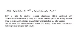 Glutathione S-Transferase Assay Kit
-
 Microscale Reduced Glutathione assay kit
-
 Glutathione Reductase Activity Coefficient Assay Kit
-
 Angiotensin Converting Enzyme Kit
-
 Glutathione Peroxidase (GSH-PX) Assay Kit
-
 Cloud-Clone Multiplex assay kits
| Catalog No. | Related products for research use of Rattus norvegicus (Rat) Organism species | Applications (RESEARCH USE ONLY!) |
| DSI523Ra01 | Rat Model for Cerebral Ischemia (CI) | Used to study the pathogenesis of cerebral infarction, pathophysiological process and thrombolytic therapy. |
| DSI523Ra02 | Rat Model for Cerebral Ischemia (CI) | n/a |
| DSI523Ra03 | Rat Model for Cerebral Ischemia (CI) | n/a |
| TSI523Ra01 | Rat Heart Tissue of Cerebral Ischemia (CI) | Paraffin slides for pathologic research: IHC,IF and HE,Masson and other stainings |
| TSI523Ra03 | Rat Liver Tissue of Cerebral Ischemia (CI) | Paraffin slides for pathologic research: IHC,IF and HE,Masson and other stainings |
| TSI523Ra05 | Rat Spleen Tissue of Cerebral Ischemia (CI) | Paraffin slides for pathologic research: IHC,IF and HE,Masson and other stainings |
| TSI523Ra07 | Rat Lung Tissue of Cerebral Ischemia (CI) | Paraffin slides for pathologic research: IHC,IF and HE,Masson and other stainings |
| TSI523Ra09 | Rat Kidney Tissue of Cerebral Ischemia (CI) | Paraffin slides for pathologic research: IHC,IF and HE,Masson and other stainings |
| TSI523Ra11 | Rat Cerebellum Tissue of Cerebral Ischemia (CI) | Paraffin slides for pathologic research: IHC,IF and HE,Masson and other stainings |
| TSI523Ra13 | Rat Brain Stem Tissue of Cerebral Ischemia (CI) | Paraffin slides for pathologic research: IHC,IF and HE,Masson and other stainings |
| TSI523Ra15 | Rat Brain Tissue of Cerebral Ischemia (CI) | Paraffin slides for pathologic research: IHC,IF and HE,Masson and other stainings |
| TSI523Ra17 | Rat Small Intestine Tissue of Cerebral Ischemia (CI) | Paraffin slides for pathologic research: IHC,IF and HE,Masson and other stainings |
| TSI523Ra21 | Rat Pancreas Tissue of Cerebral Ischemia (CI) | Paraffin slides for pathologic research: IHC,IF and HE,Masson and other stainings |
| TSI523Ra35 | Rat Spinal Cord Tissue of Cerebral Ischemia (CI) | Paraffin slides for pathologic research: IHC,IF and HE,Nissl and other stainings |
| TSI523Ra41 | Rat Aorta Tissue of Cerebral Ischemia (CI) | Paraffin slides for pathologic research: IHC,IF and HE,Masson and other stainings |